
Many healthcare providers choose an MRI scan to take a deeper look at areas of the brain that may indicate the early signs of a brain tumor.
A brain MRI is not only an excellent tool for finding potential tumors, but also for determining a tumor’s size and shape, as well as for understanding the severity of a tumor.
Let’s look at how an MRI scan can find the early signs of a brain tumor, at what you can expect from a brain MRI, and at why follow-up imaging is so important.
What is an MRI scan? How does it create detailed images of the brain?
A brain MRI scan uses powerful magnets and radio waves to create detailed pictures of the inside of your body. It works by aligning the water molecules in your body with a strong magnetic field, and then using radio waves to create signals, which a computer then turns into images.
Why is a brain MRI the preferred scan for detecting tumors at an early stage?
Because of its accuracy and sensitivity, brain MRI scans are preferred because they provide extremely high-resolution images that can reveal tiny tumors and subtle changes in brain tissue, which are important for early detection.
How an MRI helps find the early signs of a brain tumor
The earlier your healthcare provider can find a brain tumor, the more effectively they can treat your condition, before treatment becomes more challenging. An MRI can spot extremely small tumors, vastly increasing your chances for a better outcome.
How does an MRI scan show the early signs of a brain tumor?
An MRI scan helps your provider to see any unusual growths or changes in the brain that could indicate a tumor. These detailed images allow doctors to spot tumors when they are still small and easier to treat, which is what makes an MRI a helpful tool for catching problems early.
How does a brain MRI find tiny brain tumors that are hard to detect early on?
The extreme sensitivity of brain MRI scans allows them to see very small tumors that might not show up on other imaging scans. With the use of these highly accurate MRI scanners, radiologists and technologists can clearly identify these very small abnormalities or changes in brain tissue.
How can a brain MRI tell the difference between benign and malignant tumors?
Brain MRI scans can tell the difference between benign and malignant tumors based on their shape, size, appearance and how they interact with other issues around it.
Malignant tissue can show up on MRI images as a white or very light mass, whereas it would be dark in color on an ultrasound image. Contrast dye, which is a substance injected into the body before some MRI scans, enables a malignant tumor to appear more brightly on MRI scan images.
What to expect from your brain MRI scan
A brain MRI scan is pretty quick and straightforward, for most people. We’ll look at what to expect during your MRI scan, whether an MRI is safe if you have a brain tumor, and how your MRI results are interpreted.
What should I expect before, during, and after my brain MRI?
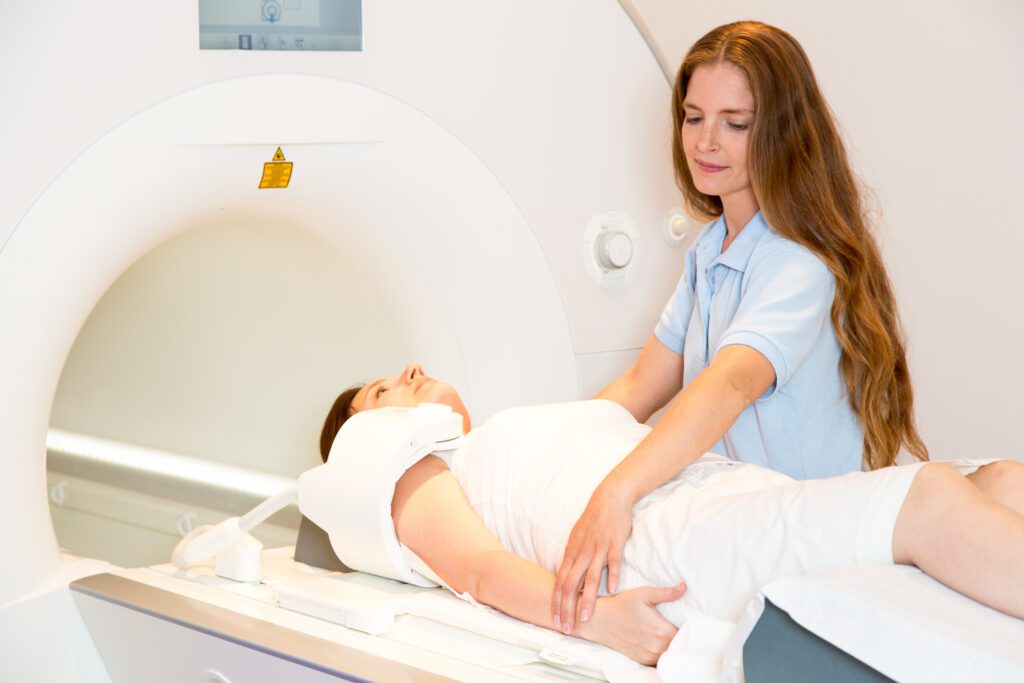
Before your brain MRI, you’ll be asked to remove any metal objects, like jewelry or watches, because the MRI machine uses a strong magnet. During the scan, you’ll lie on a table that slides into a large, tube-shaped machine. You’ll need to stay very still while the machine takes pictures, which can last about 30 to 60 minutes, depending on the scan. After the scan, you can go back to your normal activities right away, and the radiologist will review the images to help understand what’s happening in your brain.
Is it safe to get a brain MRI if I might have a brain tumor?
Yes, it is absolutely safe to have a brain MRI scan. The scan itself is non-invasive and doesn’t use any harmful radiation, which is why brain MRI scans are the preferred imaging scan for possible brain tumors or other issues
How are my MRI results analyzed? How long will it take to get my results?
After your brain MRI scan, a trained radiologist will analyze the images and will send a report to your primary healthcare provider, who will share the results with you. Touchstone Medical Imaging will also send you a text message with a link to your patient friendly radiology report. Results are typically sent 3-5 days after your brain MRI.
Why follow-up MRIs are important for monitoring benign and malignant brain tumors
If the MRI scan detects any issue, then your provider may order a follow-up MRI scan. A follow-up exam will allow your healthcare provider to compare the appearance of a tumor on your different MRI results, so they can understand it better, over time.
Why are follow-up MRIs important for tracking a brain tumor?
Your provider may order a follow-up MRI if a brain tumor was detected on your first MRI scan, to determine whether or not the brain tumor has developed further. Of course, how often you have these follow-up MRI scans will depend on your individual situation, and on your own choice.
Will I need to get follow-up MRIs for a benign brain tumor?
Depending on the advice of your radiologist and healthcare provider, if the MRI scan showed a benign brain tumor, they may want you to have another MRI scan just to make sure. Be sure to check with your provider for more information.
How can follow-up MRIs supplement a treatment plan for a malignant brain tumor?
Follow-up MRIs are an important part of a treatment plan for a malignant brain tumor, as they can help assess the progress of the treatment plan, so your healthcare team can make any changes as needed.
How to schedule your brain MRI appointment with us
Touchstone Medical Imaging offers MRI scans in Arkansas, Colorado, Florida, Montana, Oklahoma, and Texas.
Reach out to us at Touchstone Medical Imaging, and we’ll help you schedule an MRI appointment at an imaging center near you, today.
We’re here to help you get the answers you need.
FAQ's about brain MRIs for tumor detection
An MRI scan uses strong magnetic fields and radio waves to produce detailed images of the brain’s structure, helping healthcare providers see any abnormalities.
A brain MRI is preferred because it provides high-resolution images that can reveal tiny tumors and subtle changes in brain tissue, which are crucial for early detection.
An MRI can detect early signs of a brain tumor by highlighting abnormal growths, changes in tissue, or other indicators that might not be visible with other imaging methods.
A brain MRI is highly sensitive, allowing it to detect very small tumors that might be missed by other types of scans.
A brain MRI can distinguish between benign and malignant tumors based on their appearance, location, and how they interact with surrounding brain tissues.
During your brain MRI, you’ll lie still on a table that moves slowly into the doughnut-shaped MRI machine, and it will take detailed pictures of your brain.
Yes, an MRI is a safe and non-invasive procedure that doesn’t use harmful radiation, making it a reliable choice for detecting brain tumors.
Your MRI results will be analyzed by a radiologist, and you can usually expect to receive your results within a week, depending on your needs and on the imaging center.